
-
Cell & Tissue Culture
- TC Flasks
- Suspension and Shakers Flasks
- Mass Cell Culture
- 3D Cell Culture
- Dishes
- 96 & Half Area Plates
- 1-4-6-12-24-48 Wells Plates
- 384 & 1536 Wells Plates
- Advanced TC Vessels
- Protein Coated Vessels
- Cell Repellent Vessels
- TC Inserts
- Culture Tubes
- Cryogenic
- Cell Scrapers
- Cell Strainers
- CELLreactor™ Filter Top Tube
- Roller Bottles & Apparatus
- Plate Sealers & Lids
- Plant Culture Vessels
- Microplates & Sealers
- Tissue Culture Glassware
-
HTS Microplates & Sealers
- 96 Well Microplates
- 96 Wells Black & White
- 384 Wells
- 1536 Wells Microplates
- UV Plates
- 96 Wells Polypropylene Plates
- Polypropylene Storage Plates
- Sample Storage
- Glass Bottom Plates
- Non Binding Microplates
- Streptavidin Coated Microplates
- Plate Sealers
- Heat Sealable Seals
- Seal Removal Tape
- Lids and CapMats
- Magnetic Beads Acces.
- Immunology
- Bacteriology
- Molecular Biology
- Dialysis Membranes
- Chemical Labs
- Filtration & Separation
- Protein Crystallization
- Liquid handling
- Histology & Microscopy
- Glassware
- Safety Products
- Drosophlia Vials & Bottles
- General Laboratory Items
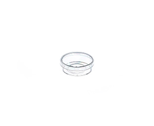
Glass Bottom Dishes

| Description | |
|---|---|
| Item No.: | 627860 |
| Product description: |
CELLVIEW CELL CULTURE DISH, PS, 35/10 MM, GLASS BOTTOM, 1 COMPARTMENT, TC, STERILE, 10 PCS./BAG |
| Packaging weight: | 0,34 kg |
| Packaging dimension: | 195 x 145 x 125 mm |
| Packing unit: | 40 |
| Quantity per inner pack: | 10 |
| Sterile: | sterile |
Detailed information
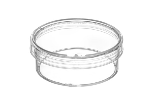
CELLview™ - Cell Culture Dish with Glass Bottom
- Free of detectable DNase, RNase, human DNA
- Non-pyrogenic, non-cytotoxic
- Glass bottom features:
borosilicate glass; hydrolytic
class 1 (DIN ISO 719)
- Advantages:
simultaneous multiplex analysis
planarity
- Number of compartments: 1
- Diameter: 35 mm; height: 10 mm
- Growth area: 8.7 cm²
- Total volume: 10 ml
- Working volume: 5 ml
- Surface treatment: TC
- Sterile
Quantity per bag/case: 10/40
Videos
Drug treatment during live cell imaging
A multi-position time-lapse experiment was started and after acquiring six time points every two minutes drugs were added to the different wells as indicated:
- Video 1 - control (no drugs added)
In steady-state the Golgi apparatus is relatively stable on light microscopy level. The shape changes only slowly during the time of the experiment when observing control cells. Also the number of Golgi fragments visible by light microscopy resolution is relatively constant over time. - Video 2 - Nocodazole added, final concentration 10 µM
Nocodazole treatment induces, fragmentation of the Golgi apparatus. The onset of fragmentation starts 10 to 15 minutes after addition of the drug. The onset of fragmentation differs between individual cells. Fragmentation of the central Golgi to many distributed ministacks is the final phenotype of microtubule depolymerization after three hours. - Video 3 - Latrunculin B added, final concentration 1 μM
Actin depolymerization by Latrunculin B influences the shape of the Golgi from relatively thin elongated to a rounded up and compact appearance. After 10 to 20 minutes differences in the Golgi morphology became first visible and after approximately one hour the Golgi
rearrangement was completed. - Video 4 - Brefeldin A added, final concentration 5 μg/ml
Block of export from the endoplasmatic reticulum (ER) by Brefeldin A leads to a rapid redistribution of the Golgi compartment to the ER by retrograde transport. This effect is often completed within 5 minutes.
Performing these experiments in parallel in CELLview™ dishes with four compartments it is possible to directly compare the speed and timing of drug effects on the Golgi apparatus. Brefeldin A affects Golgi morphology much faster than Nocodazole and Latrunculin B, which both induces first changes in the range of 10-20 minutes.
ITEM AT GREINER'S WEBSITE
| Description | |
|---|---|
| Item No.: | 627861 |
| Product description: |
CELLVIEW CELL CULTURE DISH, PS, 35/10 MM, GLASS BOTTOM, 1 COMPARTMENT, STERILE, 10 PCS./BAG |
| Packaging weight: | 0,30 kg |
| Packaging dimension: | 195 x 145 x 125 mm |
| Packing unit: | 40 |
| Quantity per inner pack: | 10 |
| Sterile: | sterile |
Detailed information
CELLview™ - Cell Culture Dish with Glass Bottom
- Free of detectable DNase, RNase, human DNA
- Non-pyrogenic, non-cytotoxic
- Glass bottom features:
- High transparent achromatic
borosilicate glass; hydrolytic
class 1 (DIN ISO 719)
- Glass thickness 175 µm +/- 15 µm
- Maximal spectral transmission; no
autofluorescence
- Advantages:
-Subdivided version enables
simultaneous multiplex analysis
- Embedded glass bottom for maximal
planarity
- Number of compartments: 1
- Diameter: 35 mm; height: 10 mm
- Growth area: 8.7 cm²
- Total volume: 10 ml
- Working volume: 5 ml
- Sterile
Quantity per bag/case: 10/40
Videos
Drug treatment during live cell imaging
A multi-position time-lapse experiment was started and after acquiring six time points every two minutes drugs were added to the different wells as indicated:
- Video 1 - control (no drugs added)
In steady-state the Golgi apparatus is relatively stable on light microscopy level. The shape changes only slowly during the time of the experiment when observing control cells. Also the number of Golgi fragments visible by light microscopy resolution is relatively constant over time. - Video 2 - Nocodazole added, final concentration 10 µM
Nocodazole treatment induces, fragmentation of the Golgi apparatus. The onset of fragmentation starts 10 to 15 minutes after addition of the drug. The onset of fragmentation differs between individual cells. Fragmentation of the central Golgi to many distributed ministacks is the final phenotype of microtubule depolymerization after three hours. - Video 3 - Latrunculin B added, final concentration 1 μM
Actin depolymerization by Latrunculin B influences the shape of the Golgi from relatively thin elongated to a rounded up and compact appearance. After 10 to 20 minutes differences in the Golgi morphology became first visible and after approximately one hour the Golgi
rearrangement was completed. - Video 4 - Brefeldin A added, final concentration 5 μg/ml
Block of export from the endoplasmatic reticulum (ER) by Brefeldin A leads to a rapid redistribution of the Golgi compartment to the ER by retrograde transport. This effect is often completed within 5 minutes.
Performing these experiments in parallel in CELLview™ dishes with four compartments it is possible to directly compare the speed and timing of drug effects on the Golgi apparatus. Brefeldin A affects Golgi morphology much faster than Nocodazole and Latrunculin B, which both induces first changes in the range of 10-20 minutes.
ITEM AT GREINER'S WEBSITE

| Description | |
|---|---|
| Item No.: | 627870 |
| Product description: |
CELLVIEW CELL CULTURE DISH, PS, 35/10 MM, GLASS BOTTOM, 4 COMPARTMENTS, TC, STERILE, 10 PCS./BAG |
| Packaging weight: | 0,40 kg |
| Packaging dimension: | 195 x 142 x 122 mm |
| Packing unit: | 40 |
| Quantity per inner pack: | 10 |
| Sterile: | sterile |
Detailed information
CELLview™ - Cell Culture Dish with Glass Bottom
- Free of detectable DNase, RNase, human DNA
- Non-pyrogenic, non-cytotoxic
- Glass bottom features:
- High transparent achromatic
borosilicate glass; hydrolytic
class 1 (DIN ISO 719)
- Glass thickness 175 µm +/- 15 µm
- Maximal spectral transmission; no
autofluorescence
- Advantages:
-Subdivided version enables
simultaneous multiplex analysis
- Embedded glass bottom for maximal
planarity
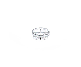
- Number of compartments: 4
- Diameter: 35 mm; height: 10 mm
- Growth area: 1.9 cm²/compartment
- Total volume: 1.5 ml/compartment
- Working volume: 0.1 ml for seeding or staining only on glass area; 0.5 ml for cultivation in the complete compartment
- Surface treatment: TC
- Sterile
Quantity per bag/case: 10/40
Drug treatment during live cell imaging
A multi-position time-lapse experiment was started and after acquiring six time points every two minutes drugs were added to the different wells as indicated:
- Video 1 - control (no drugs added)
In steady-state the Golgi apparatus is relatively stable on light microscopy level. The shape changes only slowly during the time of the experiment when observing control cells. Also the number of Golgi fragments visible by light microscopy resolution is relatively constant over time. - Video 2 - Nocodazole added, final concentration 10 µM
Nocodazole treatment induces, fragmentation of the Golgi apparatus. The onset of fragmentation starts 10 to 15 minutes after addition of the drug. The onset of fragmentation differs between individual cells. Fragmentation of the central Golgi to many distributed ministacks is the final phenotype of microtubule depolymerization after three hours. - Video 3 - Latrunculin B added, final concentration 1 μM
Actin depolymerization by Latrunculin B influences the shape of the Golgi from relatively thin elongated to a rounded up and compact appearance. After 10 to 20 minutes differences in the Golgi morphology became first visible and after approximately one hour the Golgi
rearrangement was completed. - Video 4 - Brefeldin A added, final concentration 5 μg/ml
Block of export from the endoplasmatic reticulum (ER) by Brefeldin A leads to a rapid redistribution of the Golgi compartment to the ER by retrograde transport. This effect is often completed within 5 minutes.
Performing these experiments in parallel in CELLview™ dishes with four compartments it is possible to directly compare the speed and timing of drug effects on the Golgi apparatus. Brefeldin A affects Golgi morphology much faster than Nocodazole and Latrunculin B, which both induces first changes in the range of 10-20 minutes.
ITEM AT GREINER'S WEBSITE
| Description | |
|---|---|
| Item No.: | 627871 |
| Product description: |
CELLVIEW CELL CULTURE DISH, PS, 35/10 MM, GLASS BOTTOM, 4 COMPARTMENTS, STERILE, 10 PCS./BAG |
| Packaging weight: | 0,40 kg |
| Packaging dimension: | 195 x 142 x 122 mm |
| Packing unit: | 40 |
| Quantity per inner pack: | 10 |
| Sterile: | sterile |
Detailed information
CELLview™ - Cell Culture Dish with Glass Bottom
- Free of detectable DNase, RNase, human DNA
- Non-pyrogenic, non-cytotoxic
- Glass bottom features:
- High transparent achromatic
borosilicate glass; hydrolytic
class 1 (DIN ISO 719)
- Glass thickness 175 µm +/- 15 µm
- Maximal spectral transmission; no
autofluorescence
- Advantages:
-Subdivided version enables
simultaneous multiplex analysis
- Embedded glass bottom for maximal
planarity
- Number of compartments: 4
- Diameter: 35 mm; height: 10 mm
- Growth area: 1.9 cm²/compartment
- Total volume: 1.5 ml/compartment
- Working volume: 0.1 ml for seeding or staining only on glass area; 0.5 ml for cultivation in the complete compartment
- Sterile
Quantity per bag/case: 10/40
Videos
Drug treatment during live cell imaging
A multi-position time-lapse experiment was started and after acquiring six time points every two minutes drugs were added to the different wells as indicated:
- Video 1 - control (no drugs added)
In steady-state the Golgi apparatus is relatively stable on light microscopy level. The shape changes only slowly during the time of the experiment when observing control cells. Also the number of Golgi fragments visible by light microscopy resolution is relatively constant over time. - Video 2 - Nocodazole added, final concentration 10 µM
Nocodazole treatment induces, fragmentation of the Golgi apparatus. The onset of fragmentation starts 10 to 15 minutes after addition of the drug. The onset of fragmentation differs between individual cells. Fragmentation of the central Golgi to many distributed ministacks is the final phenotype of microtubule depolymerization after three hours. - Video 3 - Latrunculin B added, final concentration 1 μM
Actin depolymerization by Latrunculin B influences the shape of the Golgi from relatively thin elongated to a rounded up and compact appearance. After 10 to 20 minutes differences in the Golgi morphology became first visible and after approximately one hour the Golgi
rearrangement was completed. - Video 4 - Brefeldin A added, final concentration 5 μg/ml
Block of export from the endoplasmatic reticulum (ER) by Brefeldin A leads to a rapid redistribution of the Golgi compartment to the ER by retrograde transport. This effect is often completed within 5 minutes.
Performing these experiments in parallel in CELLview™ dishes with four compartments it is possible to directly compare the speed and timing of drug effects on the Golgi apparatus. Brefeldin A affects Golgi morphology much faster than Nocodazole and Latrunculin B, which both induces first changes in the range of 10-20 minutes. ITEM AT GREINER'S WEBSITE
| Description | |
|---|---|
| Item No.: | 627960 |
| Product description: |
CELLVIEW CELL CULTURE DISH, PS, 35/10 MM, VENTS, ADVANCED TC, STERILE, 10 PCS./BAG |
| Packaging weight: | 2,80 kg |
| Packaging dimension: | 295 x 195 x 370 mm |
| Packing unit: | 740 |
| Quantity per inner pack: | 10 |
| Sterile: | sterile |
Detailed information
Advanced TC™ - Cell Culture Dish
- Advanced TC™ polymer modification for fastidious or sensitive adherent cells
- Free of detectable DNase, RNase, human DNA
- Non-pyrogenic, non-cytotoxic
- Advantages:
washing steps
- Improved assay consistency
- Storage at room temperature
- 2-year shelf-life
- Diameter: 35 mm; height: 10 mm
- Growth area: 8.7 cm²
- Total Volume: 10 ml
- Working volume: 5 ml
- With vents
- Sterile
Quantity per bag/case: 10/740
ITEM AT GREINER'S WEBSITE
| Description | |
|---|---|
| Item No.: | 627965 |
| Product description: |
CELLVIEW CELL CULTURE DISH, PS, 35/10 MM, GLASS BOTTOM, 1 COMPARTMENT, ADVANCED TC, STERILE, 10 PCS./BAG |
| Packaging weight: | 0,40 kg |
| Packaging dimension: | 195 x 142 x 122 mm |
| Packing unit: | 40 |
| Quantity per inner pack: | 10 |
| Sterile: | sterile |
Detailed information
CELLview™ - Cell Culture Dish with Glass Bottom
- Free of detectable DNase, RNase, human DNA
- Non-pyrogenic, non-cytotoxic
- Glass bottom features:
- High transparent achromatic
borosilicate glass; hydrolytic
class 1 (DIN ISO 719)
- Glass thickness 175 µm +/- 15 µm
- Maximal spectral transmission; no
autofluorescence
- Advantages:
-Subdivided version enables
simultaneous multiplex analysis
- Embedded glass bottom for maximal
planarity
- Number of compartments: 1
- Diameter: 35 mm; height: 10 mm
- Growth area: 8.7 cm²
- Total volume: 10 ml
- Working volume: 5 ml
- Surface treatment: Advanced TC™
- Sterile
Quantity per bag/case: 10/40
Videos
Drug treatment during live cell imaging
A multi-position time-lapse experiment was started and after acquiring six time points every two minutes drugs were added to the different wells as indicated:
- Video 1 - control (no drugs added)
In steady-state the Golgi apparatus is relatively stable on light microscopy level. The shape changes only slowly during the time of the experiment when observing control cells. Also the number of Golgi fragments visible by light microscopy resolution is relatively constant over time. - Video 2 - Nocodazole added, final concentration 10 µM
Nocodazole treatment induces, fragmentation of the Golgi apparatus. The onset of fragmentation starts 10 to 15 minutes after addition of the drug. The onset of fragmentation differs between individual cells. Fragmentation of the central Golgi to many distributed ministacks is the final phenotype of microtubule depolymerization after three hours. - Video 3 - Latrunculin B added, final concentration 1 μM
Actin depolymerization by Latrunculin B influences the shape of the Golgi from relatively thin elongated to a rounded up and compact appearance. After 10 to 20 minutes differences in the Golgi morphology became first visible and after approximately one hour the Golgi
rearrangement was completed. - Video 4 - Brefeldin A added, final concentration 5 μg/ml
Block of export from the endoplasmatic reticulum (ER) by Brefeldin A leads to a rapid redistribution of the Golgi compartment to the ER by retrograde transport. This effect is often completed within 5 minutes.
Performing these experiments in parallel in CELLview™ dishes with four compartments it is possible to directly compare the speed and timing of drug effects on the Golgi apparatus. Brefeldin A affects Golgi morphology much faster than Nocodazole and Latrunculin B, which both induces first changes in the range of 10-20 minutes. ITEM AT GREINER'S WEBSITE

| Description | |
|---|---|
| Item No.: | 627975 |
| Product description: |
CELLVIEW CELL CULTURE DISH, PS, 35/10 MM, GLASS BOTTOM, 4 COMPARTMENTS, ADVANCED TC, STERILE, 10 PCS./BAG |
| Packaging weight: | 0,40 kg |
| Packaging dimension: | 195 x 142 x 122 mm |
| Packing unit: | 40 |
| Quantity per inner pack: | 10 |
| Sterile: | sterile |
Detailed information
CELLview™ - Cell Culture Dish with Glass Bottom
- Free of detectable DNase, RNase, human DNA
- Non-pyrogenic, non-cytotoxic
- Glass bottom features:
- High transparent achromatic
borosilicate glass; hydrolytic
class 1 (DIN ISO 719)
- Glass thickness 175 µm +/- 15 µm
- Maximal spectral transmission; no
autofluorescence
- Advantages:
-Subdivided version enables
simultaneous multiplex analysis
- Embedded glass bottom for maximal
planarity
- Number of compartments: 4
- Diameter: 35 mm; height: 10 mm
- Growth area: 1.9 cm²/compartment
- Total volume: 1.5 ml/compartment
- Working volume: 0.1 ml for seeding or staining only on glass area; 0.5 ml for cultivation in the complete compartment
- Surface treatment: Advanced TC ™
- Sterile
Quantity per bag/case: 10/40
Drug treatment during live cell imaging
A multi-position time-lapse experiment was started and after acquiring six time points every two minutes drugs were added to the different wells as indicated:
- Video 1 - control (no drugs added)
In steady-state the Golgi apparatus is relatively stable on light microscopy level. The shape changes only slowly during the time of the experiment when observing control cells. Also the number of Golgi fragments visible by light microscopy resolution is relatively constant over time. - Video 2 - Nocodazole added, final concentration 10 µM
Nocodazole treatment induces, fragmentation of the Golgi apparatus. The onset of fragmentation starts 10 to 15 minutes after addition of the drug. The onset of fragmentation differs between individual cells. Fragmentation of the central Golgi to many distributed ministacks is the final phenotype of microtubule depolymerization after three hours. - Video 3 - Latrunculin B added, final concentration 1 μM
Actin depolymerization by Latrunculin B influences the shape of the Golgi from relatively thin elongated to a rounded up and compact appearance. After 10 to 20 minutes differences in the Golgi morphology became first visible and after approximately one hour the Golgi
rearrangement was completed. - Video 4 - Brefeldin A added, final concentration 5 μg/ml
Block of export from the endoplasmatic reticulum (ER) by Brefeldin A leads to a rapid redistribution of the Golgi compartment to the ER by retrograde transport. This effect is often completed within 5 minutes.
Performing these experiments in parallel in CELLview™ dishes with four compartments it is possible to directly compare the speed and timing of drug effects on the Golgi apparatus. Brefeldin A affects Golgi morphology much faster than Nocodazole and Latrunculin B, which both induces first changes in the range of 10-20 minutes.
ITEM AT GREINER'S WEBSITE
הזמנת לקוחות רשומים
לקבלת טופס על המסך יש להקיש את מספר הלקוח שלך בחברת דה גרוט בע"מ
הזמנות ישירות - כללית שרותי בריאות
:לקבלת טופס הזמנה על המסך יש להקיש את הנתונים הבאים
contact us
We are readily availbale to get your inquiry: